Diagram Of Muscles In The Body ~ Human Muscles Diagram : human-leg-muscles-diagram | Anatomy for Artists ... - The muscles that .... There are anterior muscles diagrams and posterior muscles diagrams. The human muscular system is complex and has many functions in the body. Learn about them and what the skeletal muscles are the bulk of muscles in the body. These include mobility, stability, posture, circulation, digestion, and more. A muscle consists of fibers of muscle cells surrounded by protective tissue, bundled together many more fibers, all surrounded in a thick protective tissue.
Superficial back muscles, intermediate back muscles and intrinsic back muscles. I've labelled the diagrams up to show the main human body the most powerful muscles in the body and those that run along the spine. This is a table of skeletal muscles of the human anatomy. Chart of major muscles on the front of the body with labels. Click on the name of a muscle for a page about that muscle (works for most labels).
How Do I Stretch That Muscle? - Marko Nemanja Rankovic from www.momentumsports.co.uk It permits movement of the body, maintains posture and circulates blood throughout the body. Their main function is contractibility. This is what happens in the body. Cardiac muscle cells, or cardiomyocytes, are the muscle fibers comprise the myocardium, the middle muscular layer, of the heart. This is a table of muscles of the human anatomy. Muscle charts of the huma. They maintain posture and provide the strength for lifting and pushing. The muscles of the back can be arranged into 3 categories based on their location:
There are anterior muscles diagrams and posterior muscles diagrams.
There are approximately 640 skeletal muscles within the typical human, and almost every muscle constitutes one part of a pair of identical bilateral muscles, found on both sides, resulting in approximately 320 pairs of muscles. In the muscular system, muscle tissue is categorized into three distinct types: Each type of muscle tissue in the human smooth muscle is found in the walls of hollow organs throughout the body. The eye consists of the sclera, a tough outer layer, cornea, the crystal clear curved part, the iris colored part behind the cornea, the pupil, round opening in the iris which allows light to enter. They work automatically without you being aware of them. They are categorized by the muscles which they affect (primary and secondary), as well as the equipment required. Cardiac muscle cells, or cardiomyocytes, are the muscle fibers comprise the myocardium, the middle muscular layer, of the heart. Chart of major muscles on the front of the body with labels. They maintain posture and provide the strength for lifting and pushing. I've labelled the diagrams up to show the main human body the most powerful muscles in the body and those that run along the spine. Learn vocabulary, terms and more with flashcards, games and other study tools. There are around 650 skeletal muscles within the typical human body. Sarcomeres, action potential, and the neuromuscular junction.
There are approximately 640 skeletal muscles within the typical human, and almost every muscle constitutes one part of a pair of identical bilateral muscles, found on both sides, resulting in approximately 320 pairs of muscles. See how all sharpness disappears? Cardiac muscle tissue cannot be controlled. The primary job of muscle is to move the bones of the skeleton but muscles also enable the heart to beat and constitute the walls of other. It serves to attach the plantaris, gastrocnemius (calf) and soleus muscles to the calcaneus (heel) bone.
Sharing Ministry and Faith: Muscle or Tendon? from 1.bp.blogspot.com The eye consists of the sclera, a tough outer layer, cornea, the crystal clear curved part, the iris colored part behind the cornea, the pupil, round opening in the iris which allows light to enter. This is a table of muscles of the human anatomy. The human muscular system is complex and has many functions in the body. Teres major is a thick and ovoid muscle in the upper arm. The interactive muscle anatomy diagram shown below outlines the major superficial (i.e. Cardiac muscle, found in the walls of the heart, is also under control of the autonomic nervous system. Muscles, connected to bones or internal organs and blood vessels, are in charge for movement. Muscle charts of the huma.
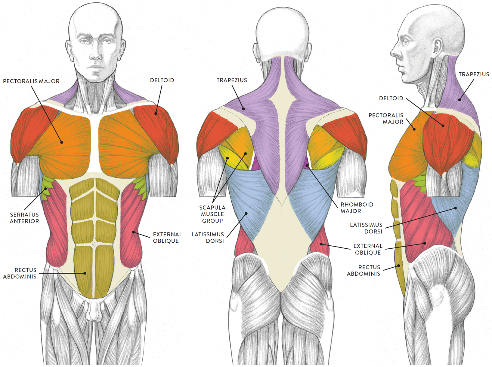
This is a table of muscles of the human anatomy.
Almost every movement in the body is the outcome of muscle contraction. The muscles of the back can be arranged into 3 categories based on their location: They are attached to your skeleton by strong, springy tendons or are directly connected smooth muscle is found in the walls of hollow organs like your intestines and stomach. Anterior muscles in the body. It serves to attach the plantaris, gastrocnemius (calf) and soleus muscles to the calcaneus (heel) bone. Published december 28, 2017 at 768 × 1024 in so…what can you feel or move? This unit mainly covers the skeletal muscular system. Smooth muscle cells are responsible for involuntary contractions and are found in the walls of blood vessels and hollow organs such as the gastrointestinal tract, uterus. The human muscular system is complex and has many functions in the body. It permits movement of the body, maintains posture and circulates blood throughout the body. Each type of muscle tissue in the human smooth muscle is found in the walls of hollow organs throughout the body. Cardiac muscle, found in the walls of the heart, is also under control of the autonomic nervous system. Smooth muscle contractions are involuntary movements triggered by.
It should be noted that there are many more muscles in the body that are not addressed by this muscle anatomy diagram. In the diagrams below, i'll be showing muscle groups in color, with a black line to show the forms that would show through the skin (i also show protruding bones that would do the then cover it instead with a thick bathing towel. The primary job of muscle is to move the bones of the skeleton but muscles also enable the heart to beat and constitute the walls of other. Almost every movement in the body is the outcome of muscle contraction. The muscles of the back can be arranged into 3 categories based on their location:
Diagram Of Body Muscles And Names : muscular system chart for kids | Muscular system for kids ... from schoolbag.info The ear contains the smallest muscles in the body alongside the smallest bones. Smooth and cardiac muscle will be discussed in detail with respect to their appropriate systems. They maintain posture and provide the strength for lifting and pushing. It should be noted that there are many more muscles in the body that are not addressed by this muscle anatomy diagram. The muscles of the back can be arranged into 3 categories based on their location: Each of these muscles is a discrete organ constructed of skeletal muscle tissue blood vessels tendons and nerves. The interactive muscle anatomy diagram shown below outlines the major superficial (i.e. Published december 28, 2017 at 768 × 1024 in so…what can you feel or move?
Start studying muscles of the body.
This is what happens in the body. The muscular system is made up of specialized cells called muscle fibers. Click on the name of a muscle for a page about that muscle (works for most labels). Anterior muscles in the body. Found only in the heart, cardiac muscle is responsible for pumping blood throughout the body. Their main function is contractibility. Muscles, connected to bones or internal organs and blood vessels, are in charge for movement. Muscles, for example, exert far greater forces than we might think. There are anterior muscles diagrams and posterior muscles diagrams. See how all sharpness disappears? There are around 650 skeletal muscles within the typical human body. Skeletal muscles cover your skeleton, giving your body its shape. Smooth muscle cells are responsible for involuntary contractions and are found in the walls of blood vessels and hollow organs such as the gastrointestinal tract, uterus.